
Fig. 1.
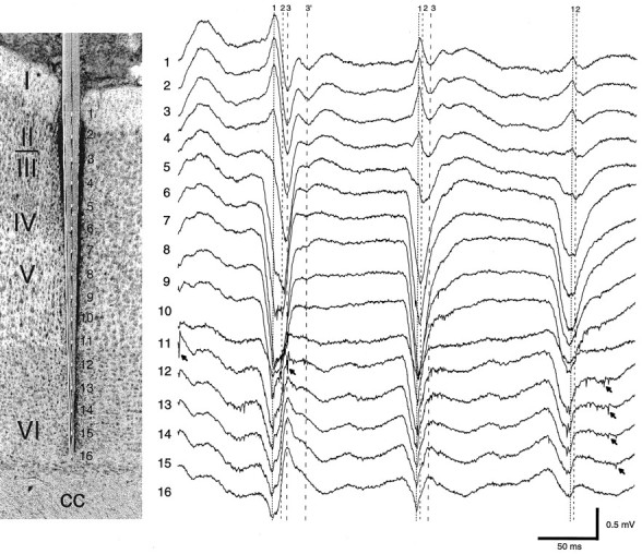
Multiple-site recording of field and unit activity in the awake rat. Histological section, The in situ location of the silicon probe and the recording sites in relation to the different cortical layers of the somatosensory area.Traces 1–16 (1 Hz–5 kHz), Three cycles of an HVS episode. Vertical lines indicate the presence of three putative dipoles contributing to the spike component of the HVS: dipole1, early surface-positive component; dipole 2, maximum negative potential in layer IV; and dipole 3, delayed surface-negative component. Dipole 3′ was occasionally observed as a separate event. Note both temporal and amplitude variation of the dipoles during successive spike-and-wave events. Arrows denote unit activity. cc, Corpus callosum.