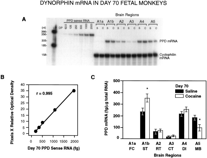
Fig. 3.
A, A representative RPA of total RNA (10 μg/lane) from saline- and cocaine-treated day 70 fetal monkey brain tissues illustrating the levels of PPD mRNA detected in the different brain regions of individual animals. UP, Undigested probe; DP, digested probe; S, saline-treated; C, cocaine-treated.A1–A5 represent defined brain regions (see Materials and Methods). B, Linear regression analysis of the optical densities of the PPD mRNA sense standard curve revealedr = 0.995. C, Distribution and quantitative analysis of PPD mRNA in brain tissue obtained from saline-treated and cocaine-treated fetal macaques (n = 3 each). Densitometric scannings were normalized to cyclophilin mRNA and quantified from each sense mRNA standard curve. PPD mRNA was significantly increased in A1b (ST and surrounding cortical regions) and significantly decreased in A5 (MB) in cocaine-treated animals (*p < 0.05; paired two-tailed Student’st test).

