
Fig. 1.
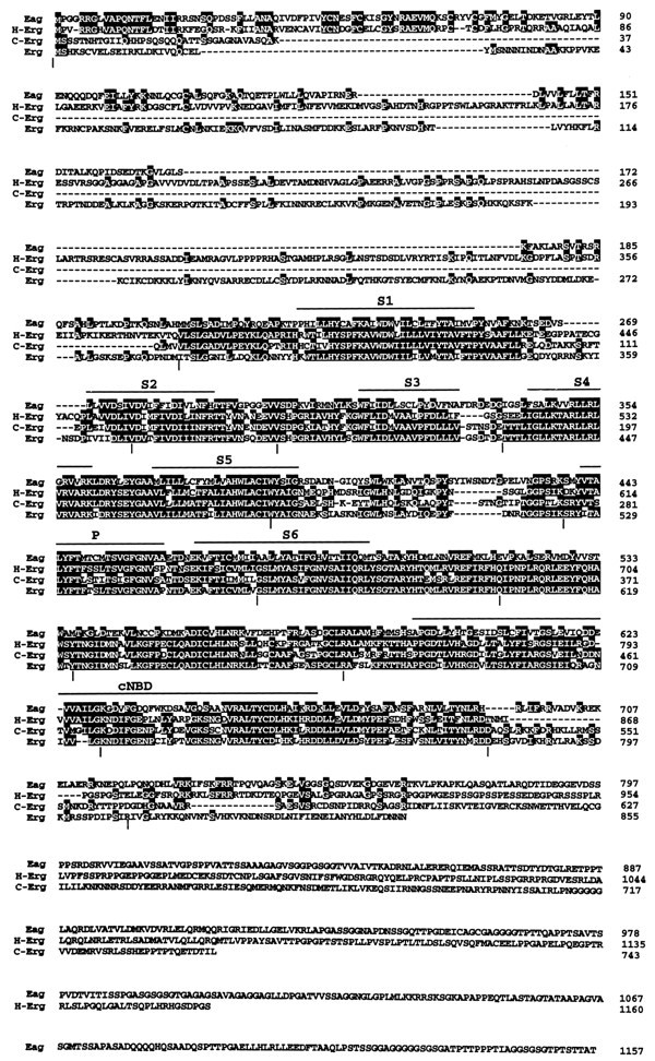
Amino acid sequence of the Drosophila erg polypeptide and its alignment with other members of theeag family of K+ channel polypeptides. Identical residues are shaded in black. The approximate locations of the presumptive membrane-spanning regions (S1–S6), the pore region (P), and the region of homology to a cyclic nucleotide-binding domain (cNBD) are overlined. Gaps in the alignment are indicated by dashes. Vertical bars beneath the alignment mark the positions of introns in theDrosophila erg genomic sequence. The Drosophila eag sequence and the human HERG sequence have been published previously (Warmke et al., 1991; Warmke and Ganetzky, 1994). The nematode erg sequence (C-erg) was obtained by using the GCG Blast program to search the GenBank database (see Materials and Methods).