
Fig. 4.
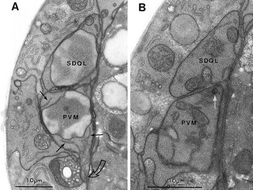
The predegeneration stage. A, Section through the somata of PVM andSDQL. In this section the cells look very similar. Note the dispersed chromatin in both nuclei (labeled with the cell names), the strands of ER in the dark cytoplasm of PVM(thin black arrows), and the characteristic ventralward process extending from PVM (open curved arrow).SDQL has a rostral process that begins to extend at this time (not shown). B, A glancing section through a more caudal portion of the same cells as in A. Thethin white arrow points to a very small circular set of nested membranes that seems to be the earliest sign ofmec-4(gf)-induced degeneration in the PVM cell. Organelles and dark cytoplasm in PVM andSDQL look similar.