
Fig. 5.
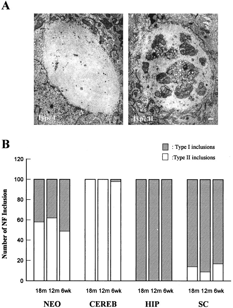
Differences in the ultrastructure of the NFHLacZ inclusions define two types of lesions. A shows representative electron photomicrographs of the two distinct types of ultrastructural morphologies of the NFHLacZ inclusions. The type I inclusion (left) is composed of compact filaments with few or no entrapped organelles. In contrast, the type II inclusion (right) entrap numerous different cellular organelles (e.g., mitochondria, endoplasmic reticulum, polyribosomes) within a tangled mass of filaments. The images in A are at the same magnification; scale bar: right, 500 nm.B shows the number of neurons with type I or type II inclusions in four different nervous regions of the TG mice at 6 weeks, 12 months, and 18 months of age. Fifty neurons from each CNS region were counted, and two animals were examined for each age group. The type I/II ratios remain quite constant in all four CNS regions throughout the life span of these mice, suggesting that the ultrastructure of the NFHLacZ inclusions is a function of the class of neuron in which it forms. NEO, Neocortical pyramidal neurons; HIP, hippocampal CA1 neurons;CERB, cerebellar Purkinje cells; SC, spinal cord motoneurons.