
Fig. 7.
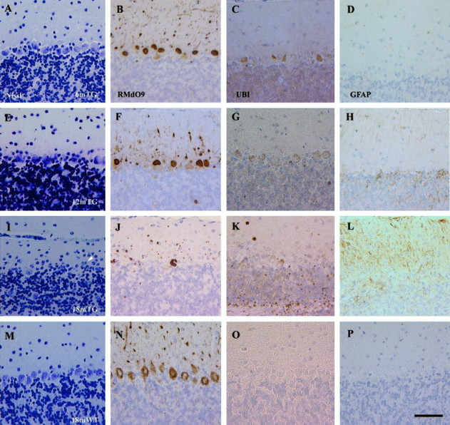
A progressive loss of Purkinje cells is detected in the NFHLacZ transgenic (TG) mice.A–D, Six-month-old NFHLacZ transgenic mouse. E–H, Twelve-month-old NFHLacZ transgenic mouse. I–L, Eighteen-month-old NFHLacZ transgenic mouse. M–P, Eighteen-month-old wild-type (WT) mouse.A, E, I, M, Nissl-stained sections. B, F,J, N, RMdO9-stained sections.C, G, K, O, Anti-ubiquitin (UBI)- stained sections.D, H, L, P, Anti-GFAP (GFAP)- stained sections. The loss of Purkinje cells starts in the 12-month-old TG mice (panels Eand F) and a prominent loss is identified in the 18-month-old NFHLacZ transgenic mice with the Nissl stain and RMdO9 (I, J). A degenerating Purkinje cell in the 18-month-old transgenic mice is shown in I(arrowhead). A concomitant increase in the immunoreactivity of anti-ubiquitin and GFAP is observed in the cerebellum of the 18-month-old NFHLacZ transgenic mice but not in the age-matched wild-type mice. The antibody-labeled sections were counterstained with hematoxylin. A–P are at same magnification; scale bar: P, 50 μm.