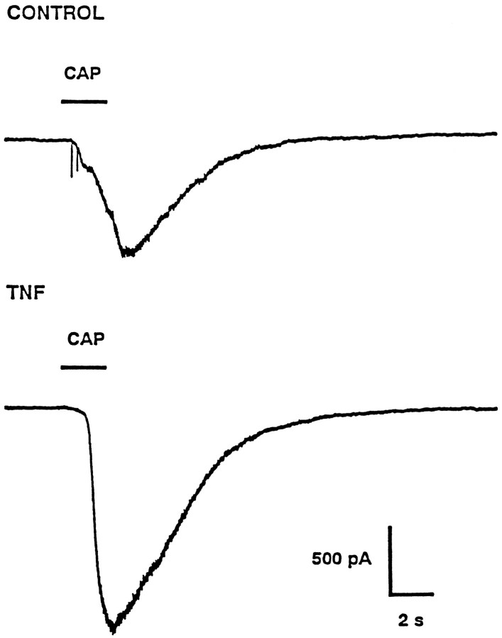
Fig. 3.
TNFα enhances the amplitude of the capsaicin response. The top panel illustrates a representative response to the focal application of 100 nm capsaicin obtained under control conditions. The bottom panelshows the response from a different neuron to the focal application of capsaicin after a 24 hr treatment with 10 ng/ml TNFα. Thebars labeled CAP indicate the timing and duration of the applications of capsaicin. Both neurons were held at −60 mV; inward currents are shown as downward.

