
Fig. 1.
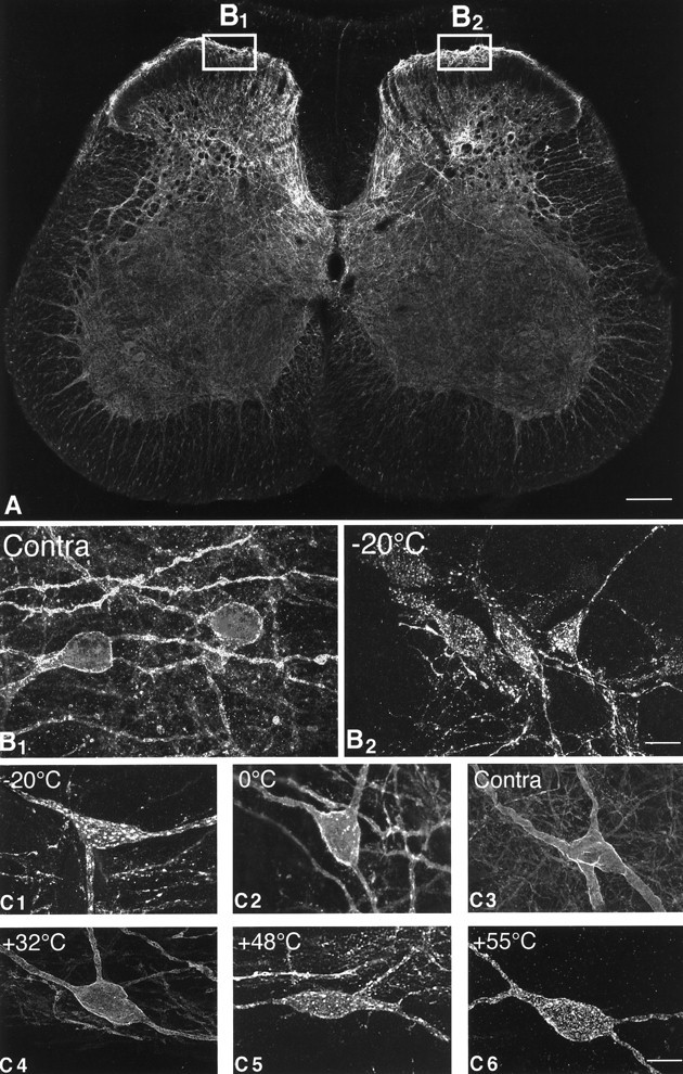
Confocal images of SPR immunoreactivity in the rat lumbar spinal cord. A, Confocal image of the rat spinal cord (L4 segment) 8 min after the plantar surface of the right hindpaw was stimulated for 30 sec with a 55°C thermode. Theboxes show the areas of the dorsal horn of the spinal cord that were sampled on the contralateral and stimulated sides. This medial aspect of the spinal cord is also the area that receives nociceptive inputs from the plantar surface of the hindpaw. InA the SPR immunoreactivity appears white, whereas in B and C panels the SPR immunoreactivity appears gray (low levels) andwhite (high levels). B1,B2, Confocal images, projected from 11 optical sections acquired at 0.7 μm intervals, of SPR immunoreactivity in the contralateral and ipsilateral sides, respectively, of the lumbar spinal cord after a −20°C stimulus to the right hindpaw. In the contralateral, unstimulated dorsal horn (B1), the SPR immunoreactivity is present on the plasma membrane, whereas on the stimulated side (B2), the SPR immunoreactivity is associated mainly with SPR+ endosomes.C1–C6, Confocal photomicrographs, projected from 22 optical sections acquired at 0.7 μm intervals, of SPR+ lamina I cell bodies 8 min after a single thermal stimulus was delivered to the hindpaw. In the contralateral control (C3) and in the ipsilateral spinal cord after the 32°C stimulus (C4), the SPR immunoreactivity is associated with the cell surface. After noxious thermal stimuli, the cell bodies experience a loss of immunoreactivity from the cell surface and an increase in the number of SPR+ endosomes in the neuronal cell body (C1, C2, C5,C6), suggesting that there is a graded release of SP from the primary afferents that is correlated with the intensity of the noxious thermal stimulation. Scale bars: A, 0.4 mm;B1–B2, 35 μm; C1–C6, 20 μm.