
Fig. 3.
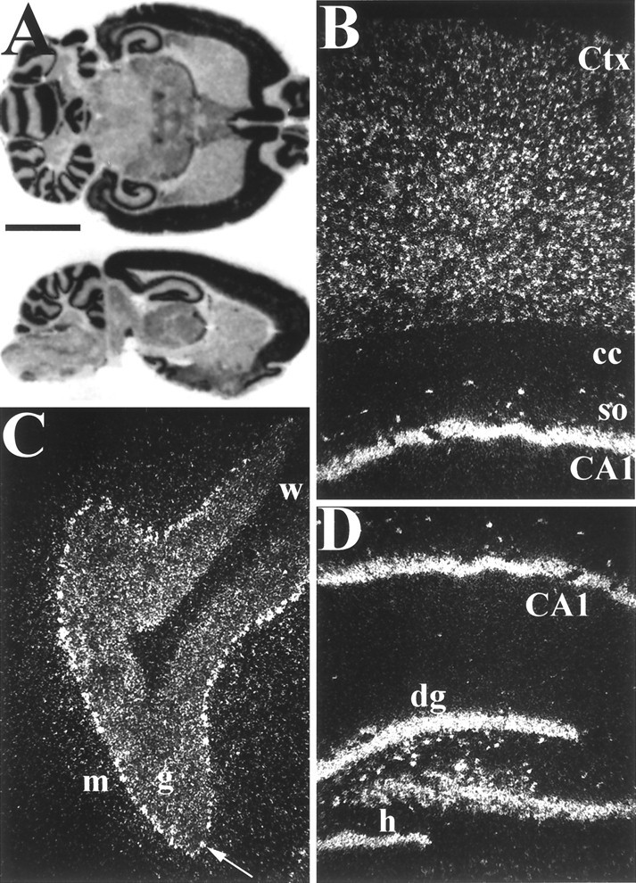
Distribution of GKAP mRNA in adult rat brain analyzed by in situ hybridization. A, Horizontal (top) and sagittal (bottom) rat brain sections probed with 35S-labeled antisense GKAP cRNA. B–D, Dark-field microscopy of sagittal in situ hybridization sections.B, Hippocampal area CA1 and overlying cortex.Ctx, Cerebral cortex, so, st. oriens;cc, corpus callosum. C, Cerebellar cortex. The arrow points to a Purkinje cell.m, Molecular layer; g, granular layer;w, white matter. D, Hippo-campal formation. dg, Dentate gyrus; h, hilus. Note prominent expression of GKAP mRNA by interneurons in the hilar region of the dentate gyrus and the st. oriens of CA1. Scale bars:A, 5 mm; B–D, 0.3 mm.