
Fig. 1.
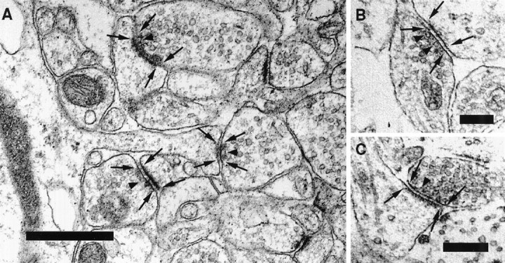
Sample electron micrographs used in our study.A, Electron micrograph depicting several synapses within the stratum radiatum in CA1 in the mouse hippocampus. Thearrows indicate the borders of the active zones on the presynaptic side and of the postsynaptic densities on the spine. Examples of vesicles that are defined as docked in our study are marked by arrowheads. B, C, Electron micrograph showing the ultrastructure of two synapses from a hippocampal neuron grown in culture. Scale bars: A, 0.5 μm; B, C, 0.25 μm.