Fig. 3.

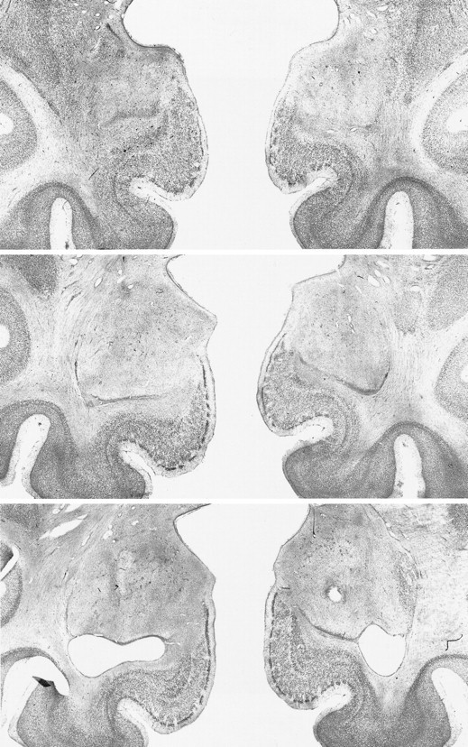
Photomicrographs of Nissl-stained coronal sections through the amygdala lesion in monkey A4. Top,middle, and bottom sections represent the left and right amygdala at approximately +18, +16, and +14 mm from the interaural plane, respectively.

Photomicrographs of Nissl-stained coronal sections through the amygdala lesion in monkey A4. Top,middle, and bottom sections represent the left and right amygdala at approximately +18, +16, and +14 mm from the interaural plane, respectively.