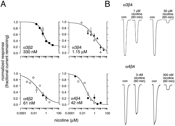
Fig. 5.
α4-containing nAChRs are desensitized by nanomolar concentrations of nicotine. A,Concentration–response plots of the ACh-induced fractional current remaining after chronic nicotine incubation in oocytes expressing α3β2, α3β4, α4β2, or α4β4 receptors. Some of the data were obtained by extrapolating the exponential fit to 60 min (α3β4 and α4β4) or 15–20 min (α3β2 and α4β2). Theopen and filled symbols represent data obtained in calcium-containing and calcium-free media, respectively. Each concentration data point represents between 1 and 10 measurements from separate oocytes. Solid lines are logistic fits to the mean of all the data obtained in calcium-free and calcium-containing media. Fits were constrained so that the maximal block could not exceed 100% and so that at infinitely low nicotine concentrations the block was zero. The half-maximal concentration for inhibition (IC50) by nicotine is shown for each nAChR subtype. B, Examples of the inhibition of the ACh test pulses by different concentrations of nicotine for two receptor subtypes, α3β4 (top traces) and α4β4 (bottom traces). Each pair of traces shows the current induced before nicotine incubation (con) and the current remaining after a 60 min incubation, with the concentration of nicotine indicated.

