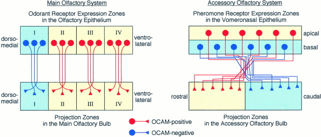
Fig. 8.
Schematic diagram of zone-to-zone projection of olfactory and vomeronasal axons in relation to zone-specific expression of OCAM. Left, In the main olfactory system four circumscribed zones (I–IV) in the dorsomedial–ventrolateral axis of the OE are defined by expression of odorant receptor genes. OCAM-positive neurons (red) are localized in the three ventrolateral zones (II–IV,yellow) of the OE and project their axons to the corresponding zones (yellow) of the MOB. OCAM-negative neurons (blue) in the most dorsomedial zone (I, light blue) send their axons to the corresponding zone (light blue) of the MOB.Right, In the accessory olfactory system the two zones,apical and basal, are stacked in the VNE as defined by the expression of G-proteins and pheromone receptor genes. OCAM-positive (red) and OCAM-negative (blue) vomeronasal neurons are localized to the apical (yellow) and basal (light blue) zones, respectively. OCAM-positive and OCAM-negative vomeronasal axons are intermingled in the vicinity of the VNE and gradually sort out to make synapses in glomeruli in the rostral (yellow) and caudal (light blue) zones of the AOB, respectively.

