
Fig. 4.
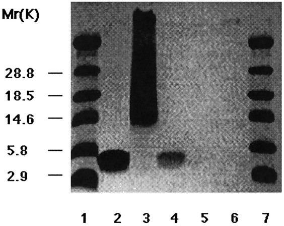
Heparin binding of apoE peptides. Peptides were incubated with heparin-agarose beads, washed, and loaded on the SDS-PAGE gel in the following order: E(141–155)2 (lane 2); E(141–149)2 (lane 3); E130–169 (lane 4); a control peptide from the C-terminal region of apoE, E268–284 (lane 5); and BSA (lane 6). Coomassie blue staining reveals that only neurotoxic apoE peptides are recovered under these conditions. Bands are present at the expected molecular weights for the peptides in addition to a smear in lane 3 attributable to aggregation of this peptide. Molecular weight markers are inlanes 1 and 7.