
Fig. 3.
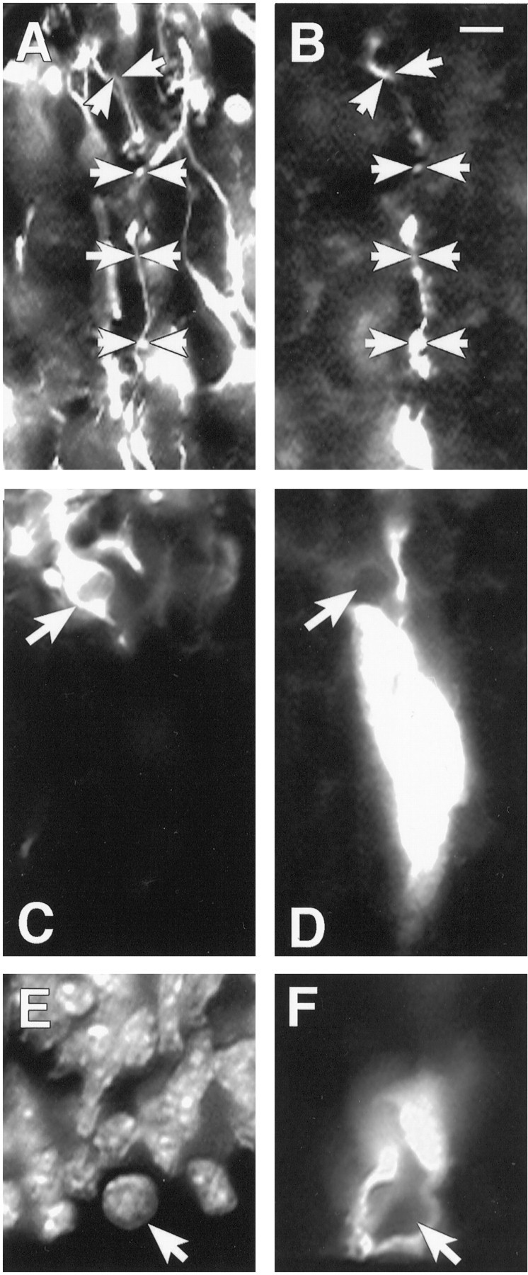
Cell types within and not within VZ clusters.A, RAT 401 nestin labeling of fibers in the IZ of E16 mouse cortex. Arrows mark the course of a glial fiber that is shown in B. B, A fiber extending into the IZ from a cluster filled in the VZ. The arrowsin A and B track the same fiber that is double-labeled for both RAT 401 and biocytin. C, TUJ labeling of the same field shown in D. Thearrow indicates a TUJ-labeled cell that is stained, and that cell is directly adjacent to the cluster labeled inD but is not coupled to the cluster. Also the fiber extending from the cluster does not stain with TUJ. D, A cluster of cells labeled with biocytin. The cluster is overexposed to show more clearly the borders of the cluster. E, A DAPI-stained view of the same field shown in F.F, The bottom of a cluster of cells at the ventricular surface. The arrow points to an M phase cell that is not coupled to the cluster. Scale bar, 10 μm.