Fig. 10.
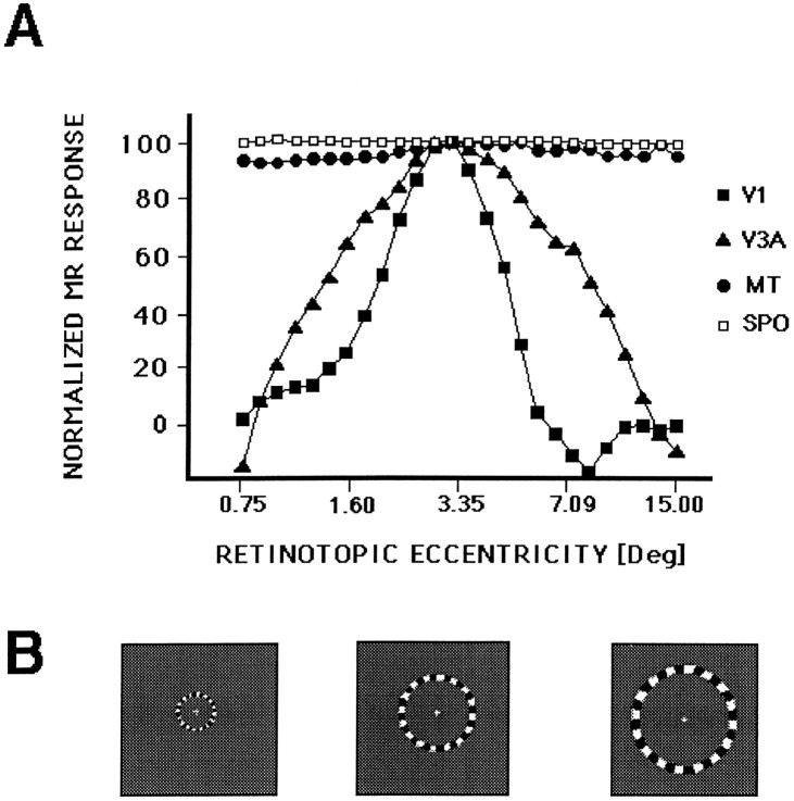
The retinotopic point spread in human V3A and comparison areas, measured as a spatiotemporal impulse function. The stimulus was a thin ring that expanded slowly and continuously (64 sec/full cycle) from the center to the periphery of the visual field, as described in Results and portrayed in B (three samples of the stimulus at different eccentricities). The fMRI time course in A was sampled from voxels in each of the visual areas, from similar eccentricities, representing the average of three consecutive scans from one subject. The borders ofV1 and V3A were defined by the field sign tests, and areas MT and SPO (the superior parieto-occipital area) were defined by motion tests (see Tootell et al., 1995a,b, 1996b) (also see Fig. 13). The retinotopic response function in V3A was consistently wider than that produced concurrently by the same stimuli in V1. The thin rings activated MT and SPO at all retinotopic locations tested, consistent with previous data showing nonretinotopic activation in these regions (Sereno et al., 1995;Tootell et al., 1995a, 1996b).

