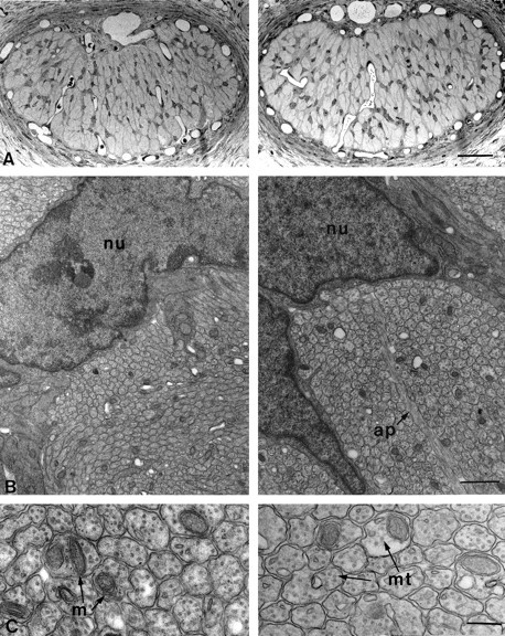
Fig. 3.

Morphology of lidocaine-treated (left) and vehicle-treated (right) optic nerves 24 hr after surgery. A, Light micrographs of semithin transverse sections of treated and control nerves in the proximity of the site of gelfoam application. There are no obvious differences between the two sections. Scale bar, 50 μm.B, Low-power electron micrographs of the specimens shown in A. Once again no qualitative differences can be seen between treated and control animals. Axons are clearly grouped in bundles separated by astrocyte processes (ap).nu, Astrocytic nuclei. Scale bar, 1 μm.C, High magnification electron micrographs showing intact, unmyelinated axons with their complement of mitochondria (m) and microtubules (mt). Scale bar, 200 nm.
