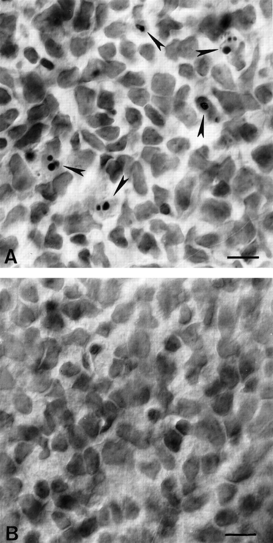
Fig. 4.

Photomicrographs of representative regions of the ganglion cell layer in whole-mounted retinae stained with cresyl violet. A, Twenty-four hours after lidocaine application onto the optic nerve, pyknotic nuclei (arrows) appear in the ganglion cell layer. B, Control retina, 24 hr after saline application. No pyknotic profiles are visible in this field. Scale bar, 10 μm.
