Fig. 5.
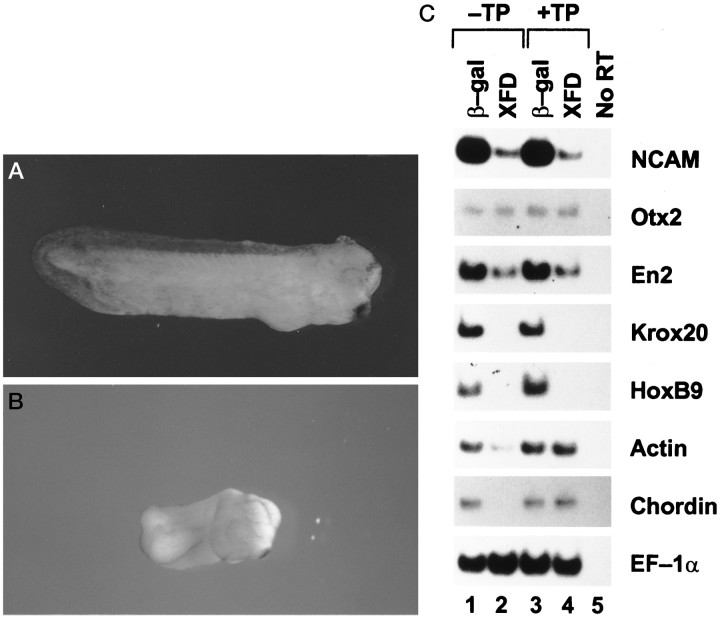
XFD inhibits posterior neural tissue in vivo. Two animal blastomeres of donor embryos were injected with 2 ng of XFD or control β-gal RNA at the two-cell stage. ACs were dissected from the injected embryos at stage 8 and transplanted to the site of neuroectoderm in recipient stage 10 embryos (+TP), which were allowed to develop to stage 30. Some XFD or β-gal RNA-injected embryos were not dissected and were allowed to develop to stage 30 as nontransplantation controls (−TP). Then, all embryos were harvested for photography (A, B) or RT-PCR analysis to detect expression of NCAM, Otx2, En2,Krox20, HoxB9, actin,chordin, and EF-1α(C). A, β-Gal and +TP;B, XFD and +TP. Photographs for XFD and −TP and for β-gal and −TP are not shown.

