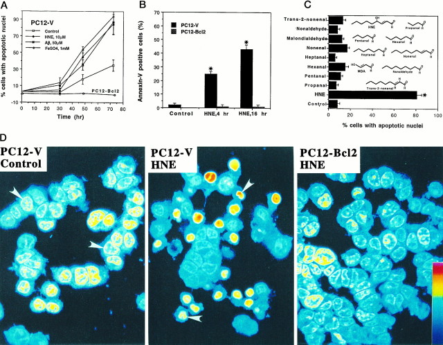
Fig. 2.
Oxidative insults and HNE induce apoptotic nuclear and plasma membrane alterations in PC12 cells: prevention by Bcl-2.A, Cultures of PC12-V cells and PC12-Bcl2 cells were exposed to vehicle (Control) or the indicated concentrations of HNE, Aβ, orFeSO4. At the indicated time points cells were stained with Hoescht dye, and the percentage of cells with condensed and fragmented (apoptotic) nuclei was determined (mean and SEM of determinations made in four separate cultures). No cells expressing Bcl-2 exhibited condensed and fragmented nuclei (PC12-Bcl2line represents combined data from cells exposed to each treatment condition). B, Annexin-V-positive cells were counted in PC12-V and PC12-Bcl2 cultures exposed for 4 hr to 10 μm HNE or for 16 hr to vehicle (Control) or 10 μm HNE. Values are the mean and SEM of determinations made in four separate cultures. *p < 0.001 compared with values for control cultures and PC12-Bcl2 cultures (ANOVA with Scheffé’spost hoc tests). C, PC12-V cell cultures were exposed for 48 hr to vehicle (Control) or the indicated aldehydes (10 μm). Cells were stained with Hoescht dye, and the percentages of cells with condensed and fragmented nuclei were determined. Values are the mean and SEM of determinations made in four separate cultures. *p < 0.001 compared with each of the other values (ANOVA with Scheffé’spost hoc tests). D, Confocal laser scanning microscope images of propidium iodide fluorescence in untreated control PC12-V cells (left), PC12-V cells exposed for 48 hr to 10 μm HNE (middle), and PC12-Bcl-2 cells exposed for 48 hr to 10 μm HNE (right). Note that many PC12-V cells treated with HNE exhibit DNA condensation and fragmentation (arrowheads), whereas cells expressing Bcl-2 did not.

