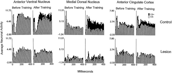
Fig. 4.
Average multiunit neuronal activity of the AV thalamic nucleus (left two columns), MD thalamic nucleus (middle two columns), and anterior cingulate cortex in response to CS+ (dark bars) and CS− (open bars) presented to rabbits with sham lesions (top row, Control) and amygdala lesions (bottom row, Lesion). The bars in each panel represent the neuronal activity in the form of z scores (see text) in 40 consecutive 10 msec intervals after the onset of the CS+ and CS−. The first, third, and fifth columnsrepresent the neuronal activity recorded during the preliminary training session in which the tones that would be used as CS+ and CS− and the foot shock were presented in an explicitly unpaired manner (neither tone predicted the foot shock). The second, fourth, and sixth columns show the neuronal activity recorded during the training session in which the acquisition criterion was attained by rabbits with sham lesions or (for rabbits that did not reach the criterion) during their last training session.

