
Fig. 2.
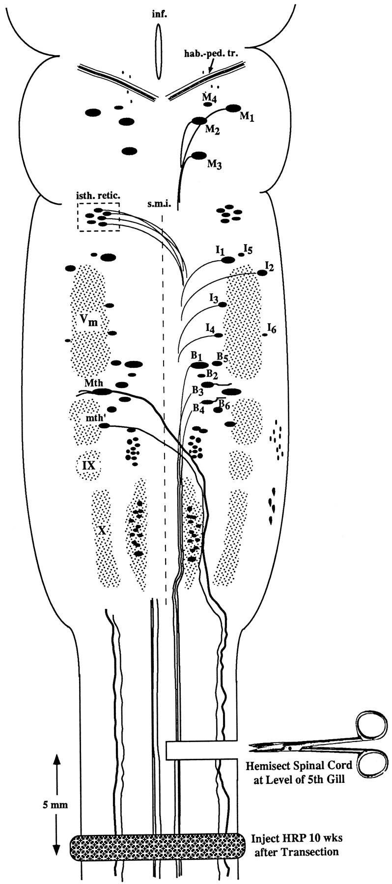
Experimental diagram of hemisection protocol. Anatomical landmarks: the brain of the large larval lamprey is diagrammed to illustrate the locations of large individually identified spinal-projecting neurons together with their mode of axonal projection (crossed or uncrossed). The left-hand members of these paired neurons are labeled according to the nomenclature of Rovainen (1967) as modified by Swain et al. (1993). M, Mesencephalic;I, isthmic; B, bulbar;Mth, Mauthner cell; mth′, auxiliary Mauthner cell; hab.-ped. tr., habenulopeduncular tract;inf., infundibulum; isth. retic., isthmic (anterior rhombencephalic) reticulospinal nucleus;s.m.i., sulcus medianus inferior;Vm, trigeminal motor nucleus; IX, glossopharyngeal motor nucleus; X, vagal motor nucleus. The unlabeled cell mass lateral to mth′ and lying between V and IX is the facial motor nucleus. Experiment: the spinal cord was hemisected on the right side at the level of the fifth gill, producing axotomy of the Müller cells and other reticulospinal neurons with uncrossed axons on the right side of the brain, and the Mauthner cell, auxiliary Mauthner cell, isthmic reticulospinal neurons, and other neurons with decussated axons on the left side of the brain. Animals were permitted to recover for 10 weeks, at which time a complete spinal cord transection was performed 5 mm caudal to the original hemisection, and HRP was applied to the cut ends of spinal cord. Previously unaxotomized neurons and neurons whose axons had regenerated were labeled by retrograde transport. Previously axotomized neurons whose axons had not regenerated at least 2.5 mm (see Materials and Methods) were not labeled.