
Fig. 3.
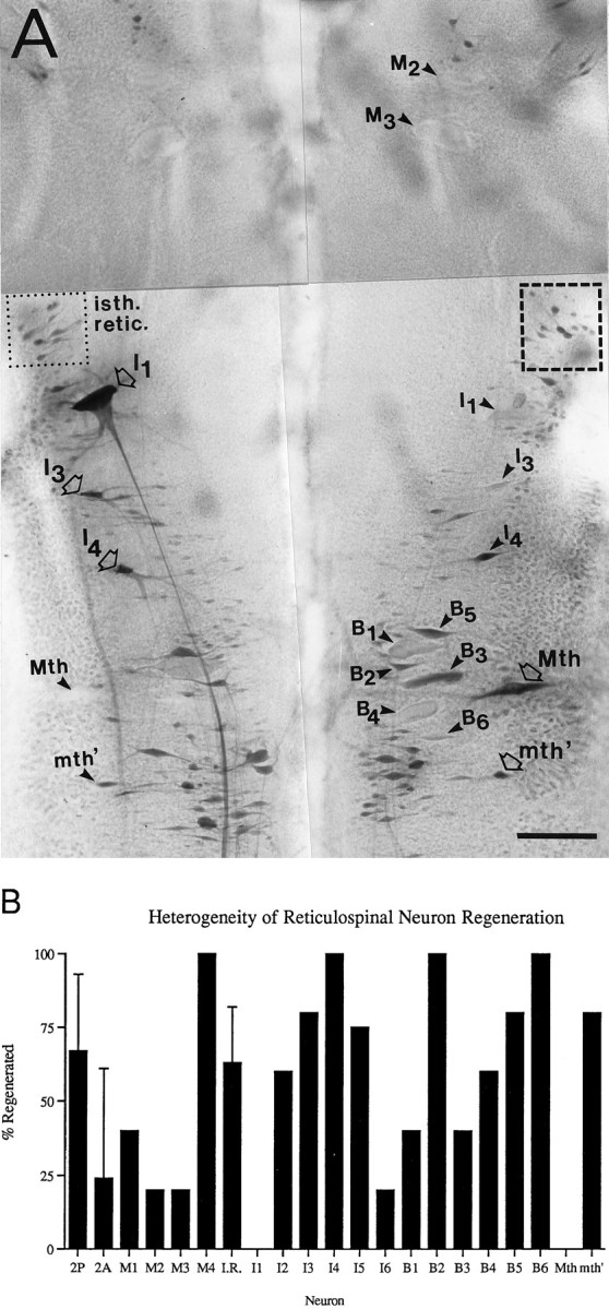
Heterogeneity in regeneration of lamprey reticulospinal axons after spinal hemisection. A, Retrograde HRP labeling of regenerated reticulospinal neurons 10 weeks after right-sided spinal hemisection. Neurons whose axons were cut by the hemisection are indicated by arrowheads, whereas neurons whose axons would not have been cut by the hemisection are indicated by open arrows. Neurons whose axons regenerated are stained darkly, as are contralateral uninjured neurons. The pale unlabeled profiles of several nonregenerating giant reticulospinal neurons are also indicated witharrowheads (e.g., M2, M3, and I1 on theright, and Mth on theleft). Numerous regenerated neurons of the isthmic reticulospinal group (isth. retic., dotted box) are stained comparably to their uninjured counterparts (dashed box). Note that axons of isthmic reticulospinal neurons and of the Mauthner (Mth) and auxiliary Mauthner (mth′) cells decussate before entering the spinal cord. Because of the close proximity of mesencephalic and some bulbar giant reticulospinal axons in the ventromedial spinal cord, contralateral axons were occasionally injured by the hemisection. This probably accounts for the lack of labeling of M1–3 on the left side of the brain. Scale bar, 150 μm. B, Probability of axonal regeneration by reticulospinal neurons 10 weeks after spinal hemisection. For each identified neuron or neuron group, the percentage of axons that regenerated was determined in five animals by retrograde filling of the cell body with HRP applied caudal to the hemisection as diagrammed in Figure 2. Standard error bars are shown for the three nuclear groups included in this figure (2P, 2A, andI.R. = isthmic reticulospinal or group 3), because these represent several neurons in each brain. These three groups were included because they were considered to have been reliably hemisected, based on control retrograde labeling experiments. It is presumed that their axons do not course near the midline at the level of the hemisection.