
Fig. 5.
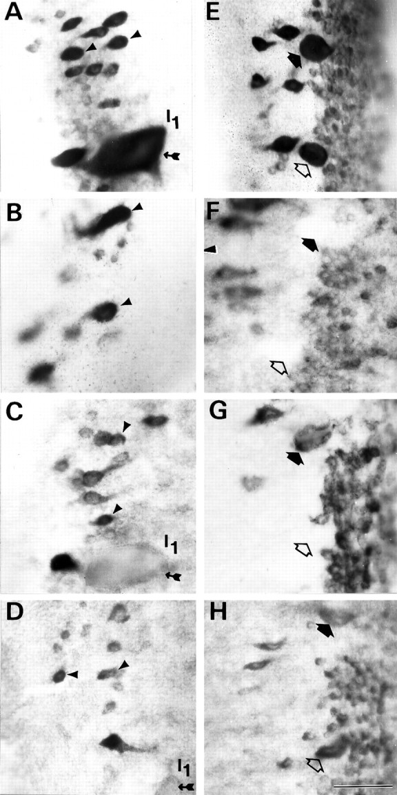
Persistence or recovery of NF-180 mRNA expression selectively in reticulospinal neurons that regenerate beyond the hemisection. The anterior rhombencephalic (isthmic) region in whole-mounted brainstems of control (A, E) and transected animals 1 week (B, F), 4 weeks (C, G), and 10 weeks (D, H) after spinal hemisection hybridized in situ to a digoxigenin-labeled NF-180 cDNA probe. Note that expression in axotomized isthmic reticulospinal neurons (arrowheads in A–D) was qualitatively reduced but remained prominent during the period when their axons were regenerating. These neurons show a high probability of axonal regeneration (see Fig. 3B; Table 1). Identified reticulospinal neurons I3 (filled arrows) and I4 (open arrows) displayed a marked early reduction in NF-180 message level that was reversed by 10 weeks (E–H). These neurons also regenerate axons effectively after transection. Note that the giant reticulospinal neuron I1 (small-tailed arrow in B–D), which almost never regenerated (Fig. 3B; Table 2), showed persistent downregulation of NF-180 expression. Scale bar, 50 μm.