Fig. 5.
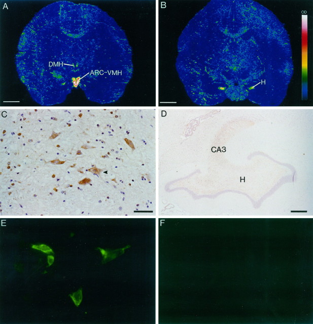
In situ hybridization autoradiograms and immunohistochemical staining of squirrel monkey brain sections and transfected HEK 293 cells showing expression of the expression of ε subunit. A, Dense mRNA expression in the arcuate–ventromedial area (ARC–VMH) and weaker expression in the dorsomedial hypothalamus (DMH). The signal intensity is indicated in the scale bar in B, with white representing the strongest signal. B, Dense mRNA expression in the hilus (H) of the dentate gyrus of the hippocampus. C, Immunoreactive cells in the DMH.Arrowhead indicates a labeled magnocellular neuron.Brown is the immunostaining reaction product, andpurple is the hematoxylin counterstain.D, Immunoreactivity in the hilus of the dentate gyrus and in the CA3 region of the hippocampus.E, ε subunit immunofluorescent labeling of HEK 293 cells transiently transfected with α1, β1, and ε subunits.F, Lack of labeling in untransfected HEK 293 cells. Scale bars: A, B, 0.5 cm;C, 10 μm; D, 100 μm.E, Magnification, 100×.

