Fig. 8.
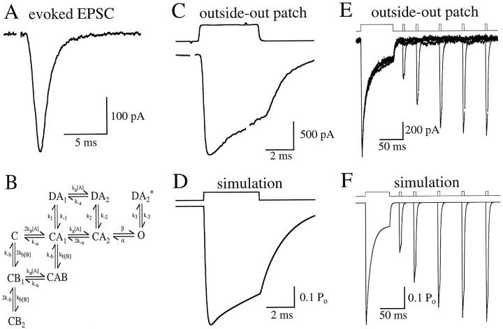
A kinetic model to mimic AMPA receptor behavior.A, Autaptically evoked AMPA receptor EPSC from a cultured CA1 hippocampal neuron (see Materials and Methods). EPSC amplitudes in this cell were unusually small. B, Markov model used to reproduce AMPA receptor kinetics observed in patch experiments. Two binding sites were configured to be equal and independent. Rates were as follows [units are μm−1 msec−1 (forka and kb) or msec−1]: ka, 0.0133; k−a, 6.24;kb, 0.03325;k−b, 5.985;k−1, 0.020;k2, 0.65;k−2, 0.018;k3, 1;k−3, 3; α, 1.1; and β, 5.7.k1 was set (0.361) to satisfy microscopic reversibility. C, l-Glu (10 mm)-evoked current in an outside-out patch excised from the same neuron as in A. The top trace shows the junction potential change caused by the solution exchange across the open tip of the electrode after patch breakdown (see Materials and Methods). In the patch response, artifacts from the voltage pulse to the piezo have been blanked. D, Simulated response to a brief 10 mm pulse of glutamate, similar to the experiment shown in C. E, l-Glu-evoked responses in an outside-out patch. Five traces are superimposed, each with a different interval (20, 50, 100, 150, and 200 msec) between the end of an initial long (70 msec) application of 10 mml-glu and the beginning of a subsequent brief (4 msec) application.F, Simulated responses to a long (50 msec) pulse ofl-glu, followed at varying intervals by brief (4 msec) pulses, similar to the experiment shown in E.

