
Fig. 4.
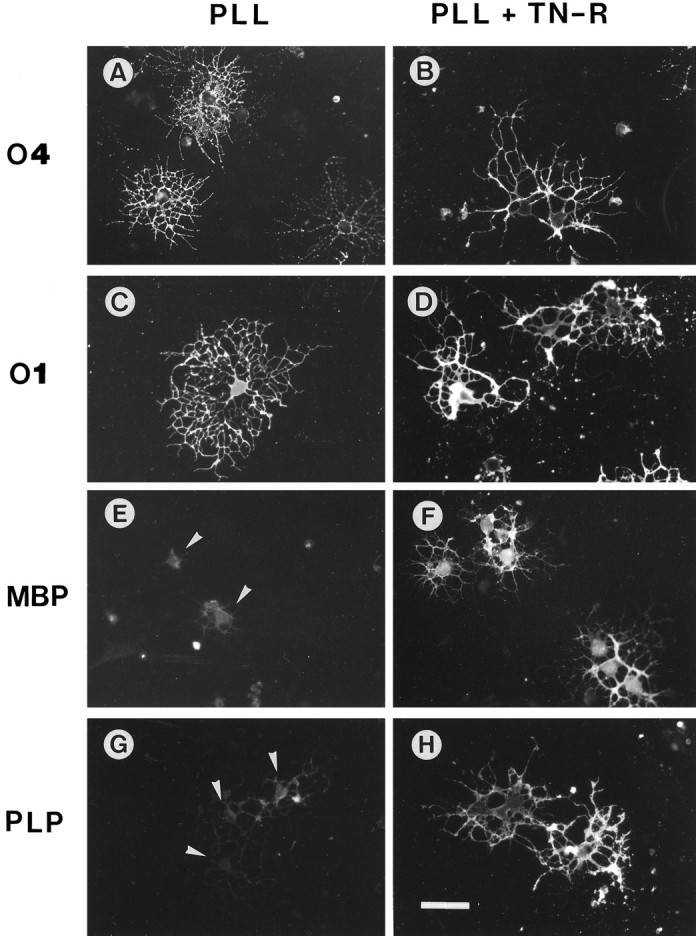
Immunocytochemistry of OL marker molecules expressed by cultured OLs in the presence of exogenous TN-R. OLs were plated onto PLL (A, C, E,G) or PLL + TN-R substrates (B, D, F, H) in modified SATO medium. The expression of O4 (A, B),O1 (C, D), MBP (E, F), and PLP (G, H) was examined after 2 d of culture by indirect immunofluorescence. Scale bar, 30 μm. Note the increase in MBP andPLP expression in the cell bodies and processes of OLs cultured on PLL + TN-R, as compared with the low myelin protein expression by OLs on PLL defined to the cytoplasm (E, G, arrowheads).