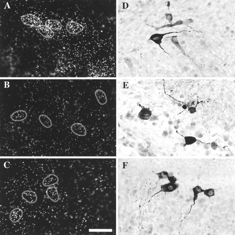
Fig. 5.

Transcription inhibitors decreased TH mRNA, but TH-immunopositive cells were still detectable. After a 40 hr exposure to transcription inhibitors (B, E, DRB;C, F, actinomycin D), TH mRNA levels decreased significantly (compare with control A), yet immunostaining revealed numerous TH-positive cells (compare with control D). All explants shown here are from the region of the CH. Circles in D–Findicate cells expressing TH mRNA. Scale bar, 50 μm.
