Fig. 1.

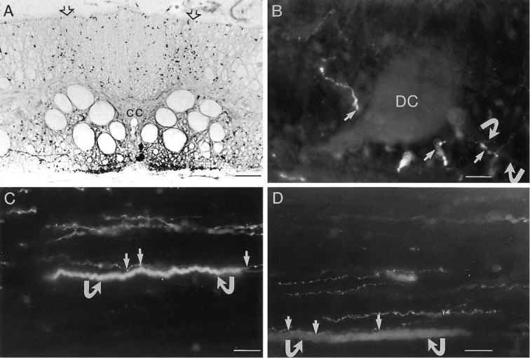
5-HT-immunoreactive (ir) fibers make close appositions with dorsal cell axons. A, A transverse section of the lamprey spinal cord showing 5-HT immunoreactivity. The dorsal column is richly innervated with 5-HT-ir fibers, which enter through dorsal roots (open arrows). Ventral to the central canal (cc), intrinsic 5-HT neurons give rise to the ventral plexus. B, A horizontal section of the spinal cord showing a dorsal cell (DC) filled with Lucifer yellow and immunoreactivity against 5-HT. Close appositions were found between 5-HT-ir varicose fibers and the cell body of the dorsal cell (arrows). Close appositions also were found on the proximal axon (curved arrows). C, D, Double-exposure photomicrographs for both Texas Red and Lucifer yellow. Dorsal cells were filled with Lucifer yellow, and the preparation was fixed and used for 5-HT immunoreactivity. 5-HT-ir fibers project in the dorsal column and make close appositions (arrows) with dorsal cell axons (curved arrows). Scale bars: A, 60 μm; B–D, 20 μm.
