
Fig. 2.
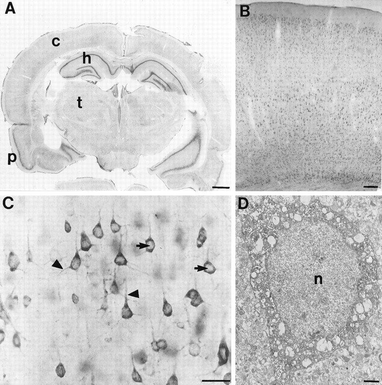
FMRP immunocytochemistry in the rat brain.A is a coronal section demonstrating widespread FMRP labeling. The most intense labeling is in the cellular layers of the hippocampus (h) and pyriform cortex (p), which are regions with extremely high neuronal densities. The deeper layers of the cerebral cortex (c) are also well labeled. B demonstrates FMRP immunoreactivity in the frontal cortex at higher magnification. It appears that most neurons in each cortical layer are FMRP-positive.C illustrates the cellular pattern of FMRP immunoreactivity in layer V pyramidal cells from frontal cortex. Staining is very dense in perikarya and proximal dendrites (triangles). In contrast, nuclear staining (arrows) is uncertain. D is an electron micrograph of the soma of a cerebral cortical pyramidal cell. With immunoperoxidase, dense cytoplasmic staining is evident. Although the nucleus is somewhat dark, FMRP immunoreactivity is not clearly present. Scale bars: A, 50 mm; B, 100 μm;C, 50 μm; D, 1 μm.