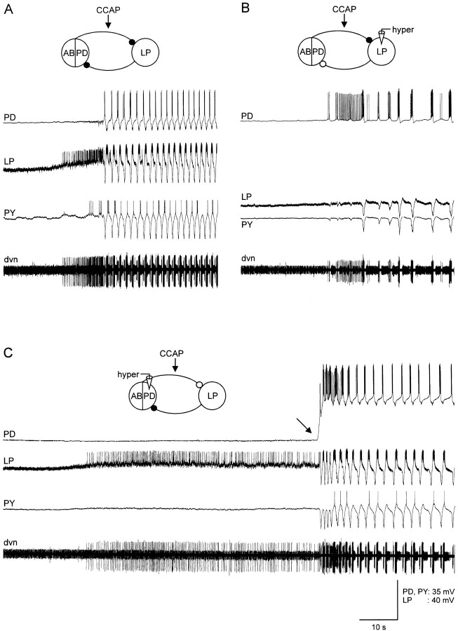
Fig. 6.
CCAP starts the pyloric rhythm in silent preparations. All panels show simultaneous intracellular recordings from the PD, LP, and PY neurons with an extracellular recording from the dnv. The electrically coupled AB and two PD neurons are shown in the drawings as a linked ensemble. The black circlesshow the chemical inhibitory synapses in control saline. Theclear circles show the synaptic connections that are presumed to be no longer active because the presynaptic neurons were hyperpolarized below their synaptic release threshold. In each panel, 10−6m CCAP was applied to the preparation shortly before the recordings shown were taken. A, Control. Initial membrane potential: PD, −54 V; LP, −63 mV; PY, −60 mV. B, LP was hyperpolarized. Membrane potentials: PD, −53 mV; LP, −100 mV; PY, −61 mV. C, PD neuron was hyperpolarized. Membrane potentials: PD, −88 mV; LP, −64 mV; PY, −60 mV.

