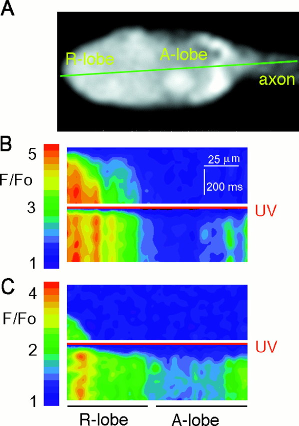
Fig. 1.

Photolysis of caged InsP3 initiates calcium release throughout Limulus ventral photoreceptors. A, Ventral photoreceptor loaded with 10 mm GDP-βS, 1 mm Fluo-3, and 10 mmcaged InsP3. The laser beam scanned along the green line every 4 msec, and the resulting lines of fluorescence data were stacked to create the images below.B, Upper frame, Fluorescence recorded during a line scan with a 488 nm laser beam, which excited the photoreceptor through the physiological mechanism. The scan shows the temporal progression of the physiological elevation of Caifrom the edge of the R-lobe membrane (left-hand edge of scan) into the A-lobe (right-hand side of scan). The laser beam started scanning across the cell at a time indicated by the first line of the image. The left-hand side of each scanned line was cropped to eliminate pixels beyond the edge of the R-lobe (determined as the point at which total fluorescence drops by 50%). The right-hand edge of each scanned line also was cropped to eliminate the final 100 pixels in which laser illumination was not uniform (see Materials and Methods). B,Lower frame, A 10 msec flash from a UV (351/364 nm) laser, relative intensity 0.5 (red line), was superimposed at the beginning of the scan. The resulting photolysis of caged InsP3 induced fast calcium release throughout the cell. C, To exclude any contribution of Ca2+influx, we kept a different cell in darkness for 30 min in ASW containing 1 mm EGTA instead of 10 mmCa2+. This increased the latency of response to the 488 nm laser via the physiological mechanism (upper frame) but did not alter the latency or pattern of elevation of Cai after photolysis of InsP3 (lower frame).
