Fig. 10.
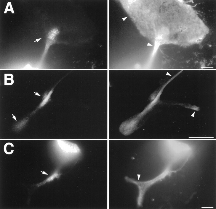
Double-labeling of bag cell neurons with antibodies to BCCa-I and ELH. BCCa-I staining is shown in theleft panel in each case, whereas corresponding staining with rat αELH is shown on the right.Arrows indicate sites of αBCCa-I staining;arrowheads, in contrast, show regions where αELH, but not αBCCa-I, staining is robust. A, A growth cone at the end of a short neurite has a broad web that stains for ELH but not BCCa-I. B, A branching neurite has prominent BCCa-I staining only at the branch-point, with less staining at the growth cone. ELH staining, in contrast, is relatively uniform.C, Accumulation of BCCa-I in a neurite is much more restricted than that of ELH, which tends to fill the neurite. The brightly stained bag cell soma is partially occluded in the top right-hand corner of each frame. Scale bars: A, C, 25 μm; B, 50 μm.

