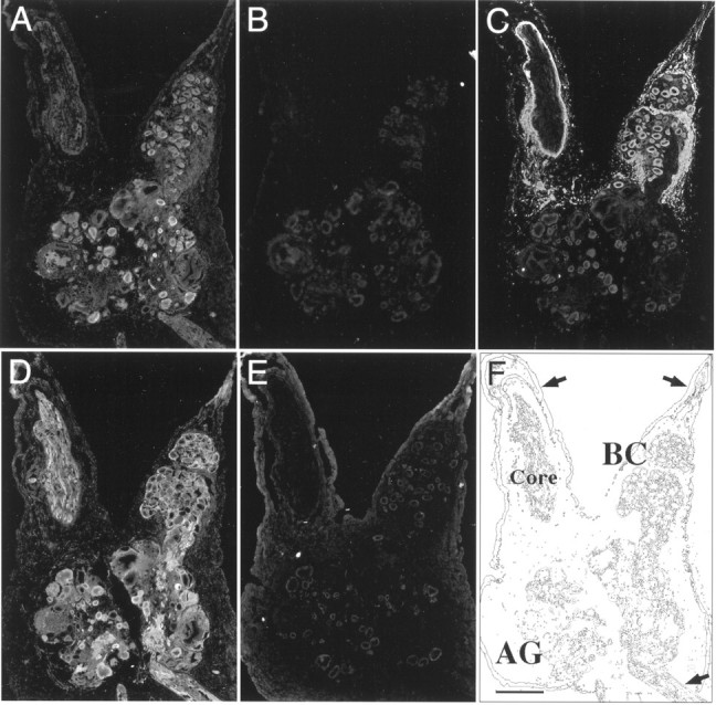
Fig. 5.

Immunofluorescent staining of tissue sections through the abdominal ganglion with αBCCa-I, αBCCa-II, and αELH antibodies. Neighboring 12 μm cryostat sections through the abdominal ganglion (AG), including one bag cell cluster (BC), were probed with antibodies to BCCa-I (A, B) and BCCa-II (D,E) and then stained with an FITC-conjugated secondary antibody. In each case, the antibodies were preadsorbed with a 100-fold molar excess of either FP-I (B, D) or FP-II (A, E) bound to glutathione-agarose beads and centrifuged at 100,000 ×g to remove bound antibody. The bag cell neurons, identified by staining with an antibody against ELH (C), are shown for comparison, and the anatomy of the tissue sections is shown schematically in F, where the arrows indicate major nerves. The top arrows indicate the pleural abdominal connectives with the axon core of the left connective labeled. The arrow on thebottom right indicates the siphon nerve. Scale bar, 500 μm.
