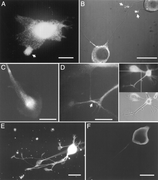
Fig. 8.
Immunofluorescent staining of bag cell neuron somata, processes, and growth cones by αBCCa-I and αBCCa-II. Cultured bag cell neurons were fixed, permeabilized, and stained for BCCa-I (A–D) and BCCa-II (E,F) as described in Materials and Methods.A, A bag cell neuron displaying the punctate pattern of BCCa-I staining. The arrow indicates an accumulation of punctate staining in one of the lammelae. Scale bar, 100 μm.B, A confocal section through the nuclei of two bag cell neurons stained for BCCa-I, overlaid on a Nomarski image of the same two neurons, and their processes. Punctate staining (inwhite) is seen cytoplasmically in the somata and in the overlapping processes (arrows). Scale bar, 100 μm.C, Process of a bag cell neuron showing the accumulation of BCCa-I staining in the growth cone. Scale bar, 50 μm.D, Accumulation of BCCa-I staining in a region of neurite contact. The top and bottom right-hand panels show the pattern of immunofluorescence and the phase-contrast image, respectively, of two bag cell neurons and their processes. The left-hand panel shows an enlarged view of the region of contact (arrow) boxed in the top right-hand panel. Scale bar, 50 μm. E, A bag cell neuron with multiple processes and growth cones showing the typically uniform pattern of BCCa-II staining. Scale bar, 50 μm.F, Confocal section through a bag cell neuron stained for BCCa-II. The highest density of staining is at the peripheral edge. Scale bar, 100 μm.

