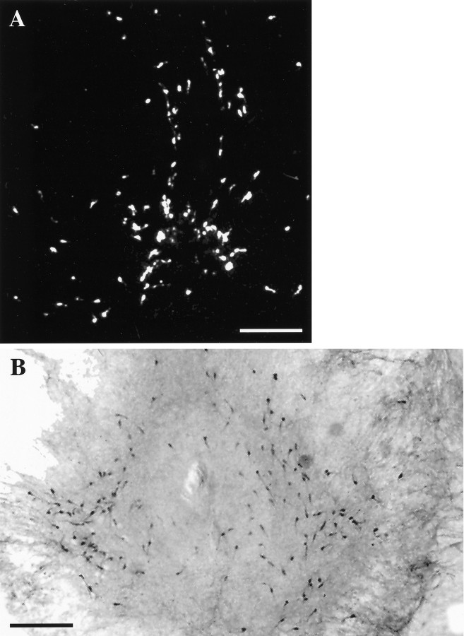
Fig. 2.
LHRH-expressing cells maintained in slice explant culture for 18 d in vitro. A, Dark-field photomicrograph of a slice 3 culture processed for ISHH, using a synthetic deoxynucleotide antisense probe for LHRH mRNA.B, Bright-field photomicrograph of a slice 4 culture immunocytochemically stained for LHRH. Note the bilateral distribution of LHRH neurons in culture. Scale bar, 500 μm.

