
Fig. 1.
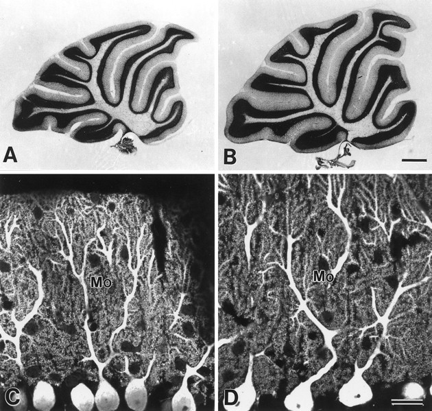
Cerebellar histology and Purkinje cell cytology in the GluRδ2 mutant (A, C) and wild-type mouse (B, D). A, B, Nissl-stained sagittal cerebellar sections. Note reductions in thickness of the granular and molecular layers in the mutant cerebellum. Rostral is to the right, and dorsal is at the top. C, D, Confocal laser scanning microscopic images of PCs immunostained for spot 35/calbindin. Note elaborate branching of PC dendrites studded with numerous spines. Mo, Molecular layer. Scale bars:B, 0.5 mm; D, 20 μm.