
Fig. 2.
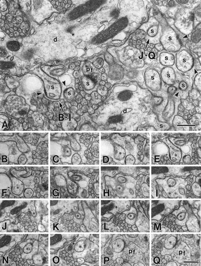
Serial electron micrographs of the molecular layer in the GluRδ2 mutant mouse at P35. A, Image from a set of serial sections. In the mutant mouse, spines (s) protruding from PC dendrites (d) occupy the molecular layer as densely as in the wild-type mouse (Fig. 3A). Note some profiles of unattached spines possess PSD-like dense materials (arrowheads) under the cell membrane.B–I, PC spine (asterisks) unattached to any nerve terminals. Note that small PSD-like dense material is seen under the postsynaptic membrane (arrowheads).J–Q, PC spine (asterisks) in contact with PF terminal (pf). Scale bars, 0.5 μm.