
Fig. 2.
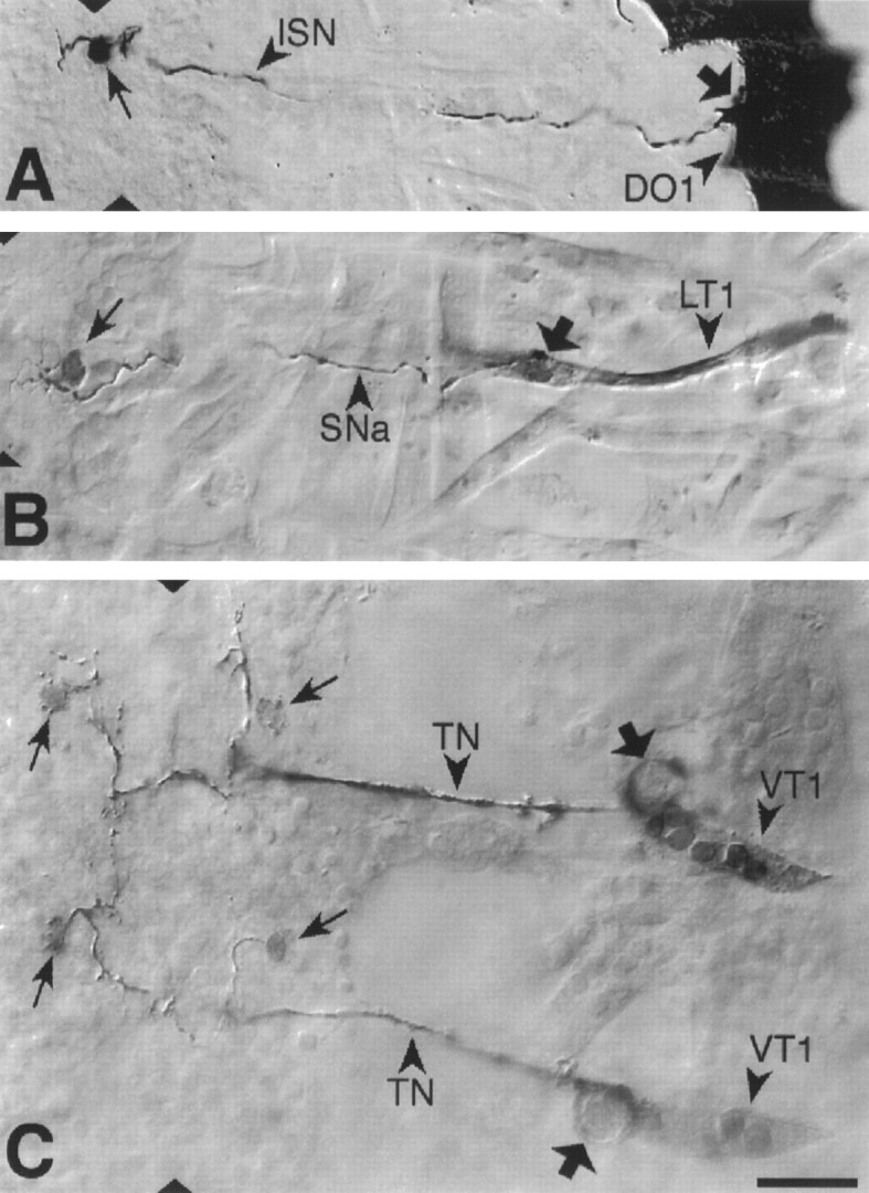
Photoconverted DiI preparations of retrogradely labeled abdominal motorneurons and their target muscles. From dorsal to ventral: (A) U motorneuron innervating muscle DO1; (B) the motorneuron that innervates muscle LT1. C, The motorneurons that innervate muscle VT1 were labeled in two adjacent segments. Arrows point to the somata and broad arrows to drops of DiI, which were deposited at the NMJs. Dorsal is right and anterior isup. The ventral midline is indicated bytriangles. Scale bar (shown in C):A, 20 mm; B–C, 10 mm.