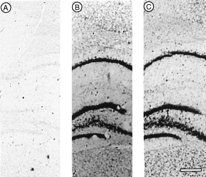
Fig. 1.
Differential expression of actin isoforms revealed by in situ hybridization. Radioactively labeled, isoform-specific oligodeoxynucleotide 33-mer probes were used to distinguish between α-smooth muscle actin (A) and the β- and γ-cytoplasmic isoforms (B, C). The autoradiograms are taken from neighboring frontal sections of rat forebrain that include (from top tobottom) the lower half of the cerebral cortex, the hippocampus (CA1, dentate gyrus, and CA4), and the upper portion of the thalamus. Whereas the α-actin probe gave only background labeling, both of the cytoplasmic actins gave strong signals in cell bodies of all areas. Scale bar, 0.4 mm.

