
Fig. 7.
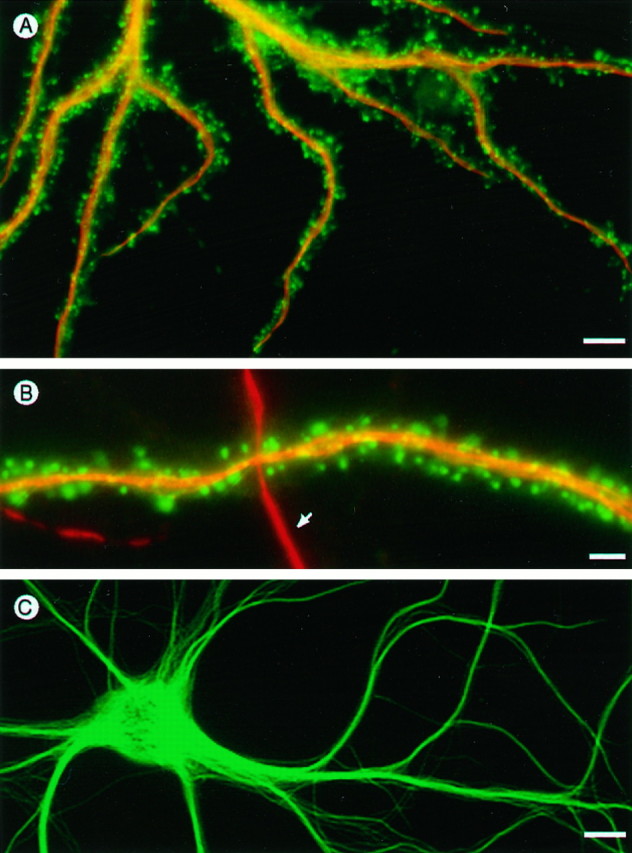
Partitioning of actin and microtubule-associated protein MAP2 in dendrites. A, Hippocampal cell transfected with vsv-tagged γ-cytoplasmic actin, fixed with glutaraldehyde, and stained with anti-vsv for actin (fluorescein channel) and MAP2 (rhodamine channel). B, A second example showing portions of a dendrite from a transfected cell (horizontal) crossed by a dendrite from a neighboring nontransfected cell (vertical, arrow). Note the contrasting pattern of γ-cytoplasmic actin in spines compared with MAP2 in the dendritic shaft domain. C, Dendrites of a living hippocampal neuron expressing GFP-tagged MAP2, which is abundant in the shaft domain but absent from dendritic spines. Scale bars: A, 10 μm; B, 1.5 μm;C, 8 μm.