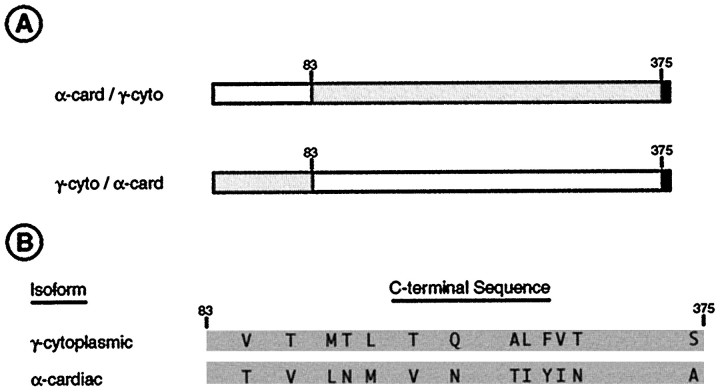
Fig. 8.
Diagrammatic summary of amino acid differences between cytoplasmic and muscle actin isoforms related to the differential targeting of actin to dendritic spines. Small numbers above the diagrams indicate residue numbers.A, Diagrams of the two chimeric isoforms used in transfection studies. γ-cytoplasmic actin sequences are shown asgray boxes, α-cardiac actin sequences by white boxes, and the vsv epitope tag by the short black box. The upper construct targets correctly to spines, whereas the lower construct does not (Fig. 6). B, Diagrams of the C-terminal portions of the γ-cytoplasmic and α-cardiac actin actin sequences with amino acid substitutions indicated at their correct relative positions.

