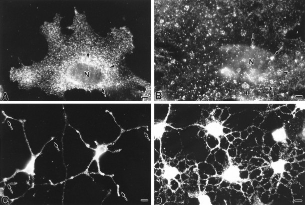
Fig. 1.
Indirect immunofluorescent localization of caveolin-1 in primary cultures of glial cells using mAb C060. A, B, The staining pattern in type 1 astrocytes is characterized by intensely fluorescent puncta that are scattered throughout the cytoplasm, particularly in the perinuclear region (arrows in A) as well as at the cell surface. At the plasma membrane, immunoreactivity is frequently organized into ring-shaped clusters (arrows inB) and is present at elevated levels at the leading or free edges of cells. C, D, Process-bearing astrocytes (C) and oligodendrocytes (D) show a prominent level of immunoreactivity at the region of the cell body. Cytoplasmic processes are decorated by fine puncta with an accumulation observed at the tips (arrows in C, D). N, Nucleus. Scale bars: A, 10 μm; B, 6 μm; C, 10 μm; D, 10 μm.

