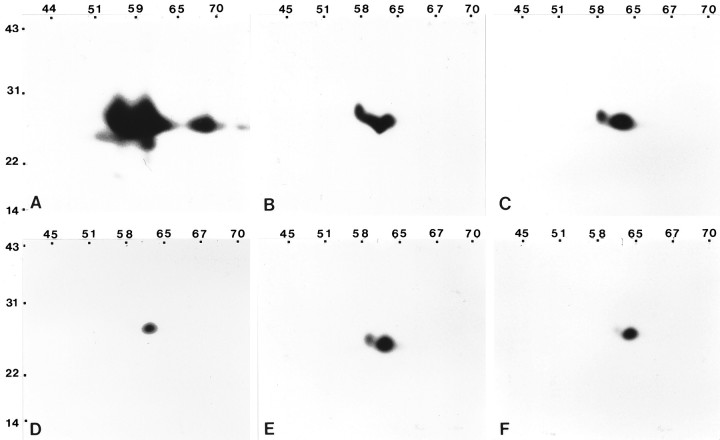
Fig. 10.
Different forms of caveolin are resolved by fractionation on Optiprep density gradients. Selected fractions were separated by two-dimensional SDS-PAGE and processed for immunoblot analyses using the anti-caveolin polyclonal antibody. Antigenic polypeptides detected in all fractions fractionate between 22 and 31 kDa. A, The most complex pattern of caveolin forms is observed in detergent-insoluble complexes isolated from the total cell homogenate. B, C, The complexity of the polypeptide pattern detected in the postnuclear supernatant (B) is simplified in comparison to that revealed by the homogenate, of which a subset is observed in the plasma membrane fraction (C). D–F, The plasma membrane fraction was sonicated and loaded at the bottom of a linear Optiprep gradient. One-milliliter fractions were collected across the gradient, and fractions 1–6, 7–11, and 12–16 were combined to form three separate pools. Each of the three pools was subjected to a second discontinuous Optiprep gradient. In each case, a single band that corresponded to the caveolin-enriched fraction was collected and processed for two-dimensional immunoblots. The spectrum of polypeptides observed for combined fractions 7–11 (E) is most similar to the spectrum of polypeptides detected in the plasma membrane fraction (C). The polypeptide profile detected for combined fractions 1–6 (D) and combined fractions 12–16 (F) appear to represent a subset of the plasma membrane (C). Molecular mass × 10−3 is indicated vertically; pH values are indicated horizontally.

