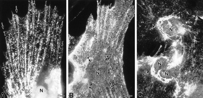
Fig. 11.
Indirect immunofluorescent localization of caveolin in primary cultures of type 1 astrocytes using affinity-purified anti-caveolin polyclonal antibodies. Primary cultures of type 1 astrocytes were fixed and permeabilized with saponin.A, B, Immunoreactivity is distributed throughout the cytoplasm in a punctate manner but is particularly evident in the perinuclear region and along linear arrays that extend from the perinuclear region to the periphery of the cell (arrowsin A and B). The linear arrays of caveolin immunoreactivity appear to have a point of origin that circumscribes the nucleus (asterisked arrows inB). C, In the perinuclear region, caveolin immunoreactivity is organized about the nucleus in a network or web-like manner (arrows in C).N, Nucleus. Scale bars: A, 6 μm;B, 5 μm; C, 7 μm.

