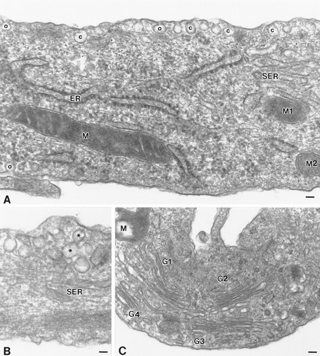
Fig. 2.

Localization of caveolae in type 1 astrocytes by electron microscopy. A, Large clusters of caveolae are distributed along the plasma membrane. Individual caveolae display a characteristic flask-shaped profile of ∼80 nm and are either open to the extracellular milieu (o) or are present as free vesicles beneath the plasma membrane (c). Frequently, the clusters of caveolae aligned along the plasma membrane appear to overlay elements of the rough endoplasmic reticulum.B, Occasionally, caveolae are detected in stacks located beneath the plasma membrane or display a figure-eight profile (*).C, The Golgi complex, although detected in abundance throughout the cell, are not typically associated with caveolae at the cell surface. ER, Endoplasmic reticulum; G1, G2, G3, G4, Golgi complex; M, M1, M2, mitochondria;SER, smooth surface tubules; o, caveolae in continuity with the plasma membrane; c, caveolae present as free vesicles beneath the cell surface. Scale bars:A, 60 nm; B, 45 nm; C, 90 nm.
