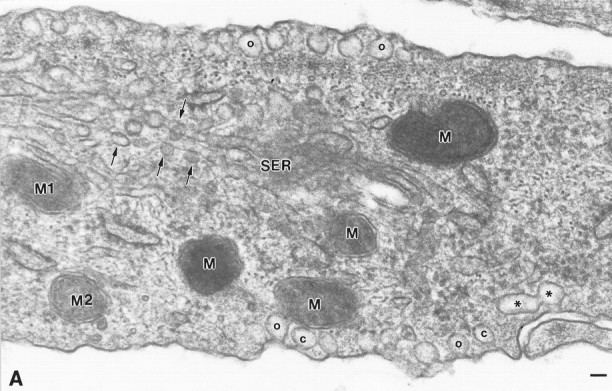
Fig. 3.

Caveolae localize to both cell surfaces in type 1 astrocytes. Coupled with Figure 2A (indicated by matching mitochondria M1 and M2), this electron micrograph reveals the great extent to which caveolae occupy both plasmalemmal surfaces. At either plasmalemmal surface, individual caveolae are either opened (o) or closed (c) to the extracellular environment. Stacks of caveolae often show figure-eight profiles (*). Intracellularly, vesicles that resemble caveolae in size and appearance are frequently observed in association with smooth surface tubules (arrows). These are most probably vesicular carriers of the exocytic pathway and likely correspond to the fluorescent puncta that are revealed throughout the cytoplasm by indirect immunofluorescence. M1, M2, Mitochondria that represent the position of overlap with Figure 2A;SER, smooth surface tubules; o, caveolae in continuity with the plasma membrane; c, caveolae present as free vesicles beneath the cell surface. Scale bar, 60 nm.
