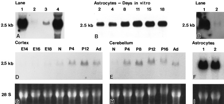
Fig. 8.
Northern blot analyses using a full-length caveolin-1 probe. Total cellular RNA was obtained as described in Materials and Methods, and 15 μg samples from cells and tissues as indicated were separated on agarose–formaldehyde gels, transferred to nylon, and hybridized with a random-primed,32P-labeled mRNA fragment for caveolin-1. Equal RNA loads were verified for all Northern analyses by ethidium bromide staining.A, A prominent band of ∼2.5 kb is detected in total RNA isolated from type 1 astrocytes (lane 1). The quantity and size of the message observed in astrocytes is identical to that detected in total RNA isolated from lung (lane 4). A band of identical size was detected in total RNA isolated from cerebellum (lane 2) and cerebral cortex (lane 3), although at significantly reduced levels.B, Astrocytes were maintained in culture from 0 to 18 d in vitro, and total cellular RNA was isolated for each time point. The level of message detected is slightly reduced for astrocytes maintained from 2 to 4 d in vitrobut thereafter remains constant. C, Total cellular RNA was obtained from primary cultures of astrocytes and from a homogeneous population of primary hippocampal neurons. The level of caveolin-1 message detected in hippocampal neurons (lane 2) is negligible in comparison to that determined for astrocytes (lane 1). D–F, Total cellular RNA was obtained at multiple developmental ages from cerebral cortex [embryonic day (E14) 14 to adult] and from cerebellum (newborn to adult). The peak level of caveolin-1 message is detected at postnatal day 12 (P12) for cerebral cortex (D) and at postnatal day 8 for cerebellum (E). The size of the message, ∼2.5 kb, is identical to that detected in total cellular RNA obtained from astrocytes (F; lanes 1, 2 represent independent astrocyte preparations). However, the level of caveolin-1 message observed is significantly reduced in brain tissues in comparison to that observed for quantitatively identical levels of RNA obtained from astrocytes. G–I, Corresponding ethidium bromide staining of the agarose–formaldehyde gel for which 15 μg samples of total cellular RNA obtained from cerebral cortex, cerebellum, and astrocytes were separated and processed for the Northern blot analyses shown in D–F.

