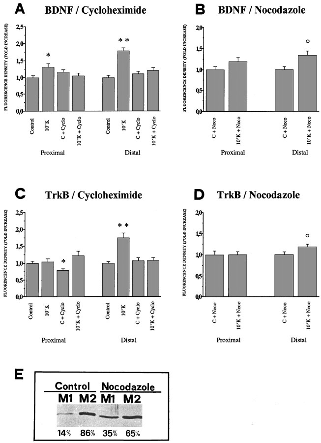
Fig. 9.
Quantitative analysis of BDNF and TrkB protein levels in proximal and distal regions of the dendrites. Immunofluorescence for BDNF or TrkB was acquired by five confocal sections and integrated in a single projection, in control conditions and after 10 min depolarization with 10 mm KCl, in the presence or absence of cycloheximide (A, C) and of nocodazole (B, D). Fluorescent density of BDNF (A, B) and TrkB (C, D) was determined in proximal and distal regions of the projections of labeled dendrites as described in Materials and Methods. Bars represent the mean fold increase of the fluorescence density of 45 dendrites, with respect to the controls (=1.0). Error bars represent SE.A, Incubation of cells in 10 mm KCl for 10 min leads to a strong increase of BDNF fluorescence density in both proximal and distal regions (10′K). Incubation of control cells with the protein synthesis inhibitor cycloheximide does not alter the basal levels of fluorescence density for BDNF in proximal and distal regions (C + Cyclo). Cycloheximide completely inhibits the increase in fluorescence density induced by the 10 mm KCl stimulus (10′K + Cyclo).B, After pretreatment of cells with nocodazole for 6 hr the levels of BDNF fluorescence density in the proximal dendrites depolarized for 10 min with 10 mm KCl in the continuous presence of nocodazole (10′K + Noco) were comparable to control (C + Noco), whereas a significant fluorescence density increase could be detected in the distal dendrites (10′K + Noco). C, Incubation of cells in 10 mm KCl for 10 min led to a strong increase of fluorescence density for TrkB in the distal region (10′K) but not in the proximal region, and this effect was abolished by cycloheximide (10′K + Cyclo). Incubation of control cells with cycloheximide reduces the basal levels of TrkB fluorescence density in the proximal but not in the distal regions (C + Cyclo). D, After pretreatment of cells with nocodazole for 6 hr, followed by a depolarization for 10 min with 10 mm KCl in continuous presence of nocodazole, the TrkB fluorescence density in the proximal dendrites (10′K + Noco) was comparable to the controls (C + Noco), whereas a fluorescence density increase could be detected in the distal dendrites (10′K + Noco). Significance with respect to controls: op ≤ 0.05; *p ≤ 0.01; **p ≤ 0.001.E, Western blot for the soluble and microtubule cellular pools of tubulin in hippocampal neurons in culture. M1, Soluble tubulin fraction; M2, polymerized tubulin fraction. After 6 hr incubation with nocodazole, the soluble, unpolymerized tubulin fraction is doubled.

