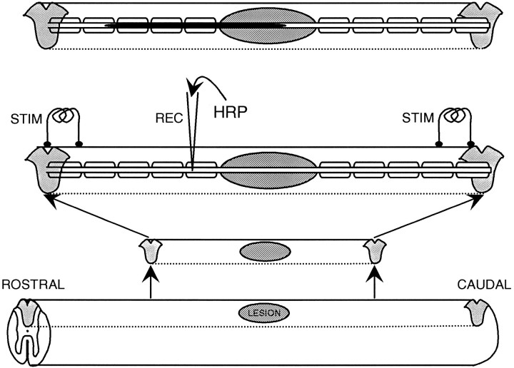
Fig. 1.
Diagram illustrating the intra-axonal electrophysiological and labeling techniques used. Starting at thebottom of the figure, the diagrams show the excision from the spinal cord of the dorsal columns containing the lesion, the arrangement of the two pairs of stimulating electrodes and the recording micropipette (which is shown inserted into an axon that is demyelinated in the lesion), and (top) the filled axon after ionophoresis of HRP. Note that the caudal pair of electrodes was also used for recording CAPs.

